Handheld Fundus Imaging for Emergency Departments and Eye Triage
Improve retinal examinations with the non-mydriatic Optomed handheld fundus camera.
Contact usTurning a Difficult Examination into a Reliable Clinical Tool
Fundus examination is an important part of neurological and emergency eye assessments.
However, performing it with traditional ophthalmoscopy can be difficult for non-ophthalmologists because:
- Success rates can be low
- The optic disc may be difficult to visualize
- Clinicians often work under time pressure
- The examination is rarely documented with images
Optomed non-mydriatic handheld fundus cameras enable reliable and fast fundus imaging and integrate smoothly into different clinical workflows in emergency, neurology and eye triage use.
Book a DemoReliable Fundus Imaging with
High Success Rate
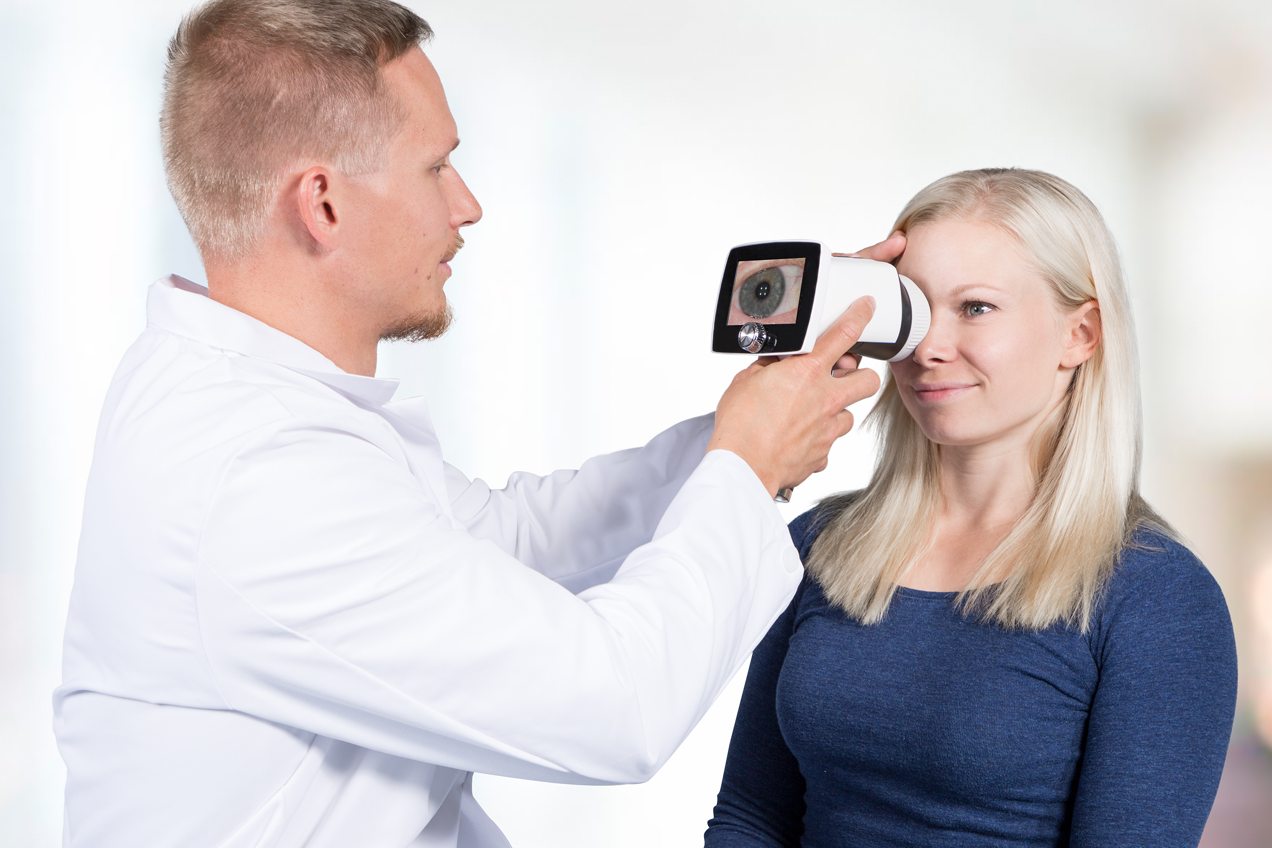
Optomed handheld fundus cameras enable fast and reliable retinal imaging in emergency and neurology settings.
Key Benefits
- High imaging success rate – 93% vs. 58%1 (compared to traditional ophthalmoscopy)
- No pupil dilation required – saves time and enables pupil reactivity monitoring
- Can be used by any healthcare staff – fundus imaging reliably performed by non-physician staff2
- Documentation with an image supports clinical decision-making and follow-up3

Clinically Validated Benefits of Optomed Fundus Imaging in the ED1
93% imaging |
Reliably completed by |
Comprehensive fundus |
Integration to |

Digital Documentation Strengthens Clinical Workflow
- Save fundus image directly into EMR / PACS
- Support remote interpretation and specialist consultation
- Enable comparisons over time for treatment progress
- Provide a fundus image as a record for medico-legal and quality assurance
Fast and Accessible Fundus Imaging at the Bedside
- Effective imaging even when the patient cannot sit up
- Non-mydriatic operation reduces exam setup time
- Digital image capture avoids repeated examinations
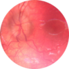
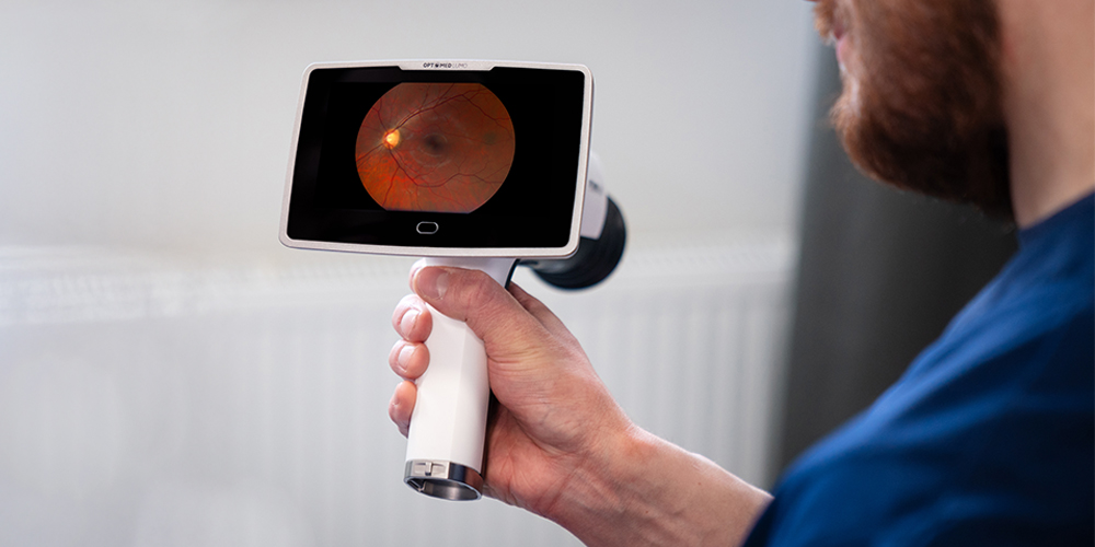
- Optic disc -centered imaging set-up with the Optomed Lumo
- Immediate image review on screen supports accurate assessment

Integrating Optomed Fundus Imaging into Real-World Clinical Workflows
Patient with headache, |
Examine the optic |
Save fundus image |
Detect presence or |
Expanded Clinical Capability with Anterior Imaging
The Optomed Aurora IQ brings versatility to the emergency department ophthalmic examinations with both anterior and posterior modules in one device.
- Capture high-quality posterior and anterior images
- Document ocular anterior segment emergencies
- Anterior segment images with color and cobalt blue filter
Read what our users think about us
The Optomed Aurora IQ is a game-changer for fundoscopic examinations in neurology
Neil Scolding, FRCP, PhD
Professor of Clinical Neurosciences Emeritus, University of Bristol Visiting Professor of Medicine, Gulu University, Uganda
I am a Consultant Neurologist and emeritus Professor of Clinical Neurosciences in Bristol, UK, but also work as a volunteer with a Visiting Professor contract at Gulu University Medical School Faculty of Medicine in northern Uganda, in undergraduate medical student teaching, post- graduate specialist training in neurology and internal medicine, clinical practice and research.
Most of our clinical work, medical student teaching and junior doctor training takes place at St Mary’s Hospital, Lacor, a non-for-profit non-government hospital which has a formal affiliation as a Teaching Hospital of Gulu University Medical School.
Teaching of ophthalmoscopic skills is never easy and the absence of ready access to MRI/CT scanning markedly increases the importance and value of good fundoscopy.
On this background, The Optomed Aurora IQ camera has been a game-changing development.
After a brief teaching lecture and a practical demonstration of the Optomed Aurora IQ, the camera was taken into regular use by junior and senior doctors in their clinical practice.
The Optomed Aurora IQ is making an enormous difference in this rather remote area of northern Uganda.
Non-mydriatic use and a large field of view good for a busy neurology clinic or ED
Dr Janice Redmond
Consultant Neurologist
St James’ Hospital Dublin
Since the arrival of the Optomed Aurora we have made use of the device for patients seen in a General Neurology setting. These patients are seen in clinic and on consult rounds throughout the hospital both on wards and in the Emergency Department. Principally we were interested in obtaining the Optomed Aurora for the examination and documentation of fundoscopy for patients with Multiple Sclerosis in clinic and those presenting to the Emergency Department and to clinic with headache disorders, in particular idiopathic intracranial hypertension.
Prior to the Optomed Aurora these patients were examined with a direct ophthalmoscope (most typically a Welch Allyn direct ophthalmoscope) which has become increasingly challenging as an examination modality in the context of COVID-19. The requirement for physicians to wear face shields and visors when examining patients has resulted in direct ophthalmoscopy being rushed or omitted in some cases. When its performance is indicated there is an unavoidable increase in the level of contact and concomitant increase in the risk of exposure. While there have been attempts made in other centres to circumvent this (Jorge, Martins and Prata, 2020), we were keen to make use of an alternative approach.
The Optomed Aurora allows for safe acquisition of retinal images while maintaining appropriate levels of contact as required during the COVID-19 pandemic. This combines with the advantage of maintaining a documentary record of fundal images which can be compared over time in our clinic patients and the capture of significant retinal findings for educational and research purposes. The wide field of view that can be obtained without the requirement for mydriatic agents renders image acquisition feasible in a busy clinic or the ED with minimal inconvenience to patient or physician.
We found the Optomed Aurora easy to use and after a single tutorial from the manufacturers we rapidly improved the speed and quality of our retinal photography. Optomed provided us with a valuable trial period which allowed us to become familiar with the camera and have given us excellent support throughout the initial use period. The set-up and operation of the camera is straightforward and easy to explain to colleagues. Moreover, the light weight and form factor makes it feasible to bring on ward rounds and to clinic.
Reference:
Jorge A, Martins AI, Prata M, et al. Ophthalmoscopy in COVID-19 low-risk patients. Practical Neurology 2020;20:425-426.
Image clarity is impressive
Prof. Dr. Tjalf Ziemssen
Center of Clinical Neuroscience, Department of Neurology, University Hospital Carl Gustav Carus, Dresden University of Technology, Germany
We have tested Aurora Camera on several healthy volunteers in the last months of 2021 before the more severe restrictions imposed by Covid pandemics. Our impression of the Aurora Camera was excellent; it is a very useful tool, especially in the emergency room and for the specialists in ophthalmology, but also in the neurovascular field for the search for vascular status. The clarity of the 2D images is impressive.
Contact us to learn more


Sources: 1) Alm M, Hautala N, Bloigu R, Huhtakangas J. Comparison of optic disc evaluation methods in neurology emergency patients. Acta Neurol Scand. 2019 Dec;140(6):449-451. 2) Mackay DD, Garza PS, Bruce BB, Newman NJ, Biousse V. The demise of direct ophthalmoscopy: A modern clinical challenge. Neurol Clin Pract. 2015;5(2):150-157. 3) Pérez MA, Bruce BB, Newman NJ, Biousse V. The use of retinal photography in nonophthalmic settings and its potential for neurology. Neurologist. 2012 Nov;18(6):350-5.