Veterinary Retinal Imaging
Optomed handheld fundus camera offers easy-to-use patented technology for veterinary eye imaging and documentation.
Contact us
Non- |

Instant |

Follow-up |

USB and |
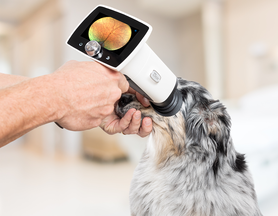
Common Uses of Fundus Imaging in Veterinary Practice
Screening for hereditary eye diseases: Document retinal findings to support breeding examinations and assessment of suspected inherited conditions.
Retinal and optic nerve examination: Examine and document retinal abnormalities, optic nerve changes and other ocular findings.
Detection and monitoring of disease: Identify ocular manifestations associated with systemic or infectious diseases and monitor changes over time.
Clinical documentation and follow-up: Capture digital fundus images for comparison, specialist consultation, owner communication and follow-up
Handheld Fundus Camera for Veterinary Retinal Imaging
Optomed Aurora IQ premium handheld fundus camera has 50° field of view and High-Contrast Optical Design™ combined with non-mydriatic operation. This allows for a detailed examination and ease of use in various cases where retinal examination is needed.
- Quick and quiet image capture
- Large screen for viewing the results instantly
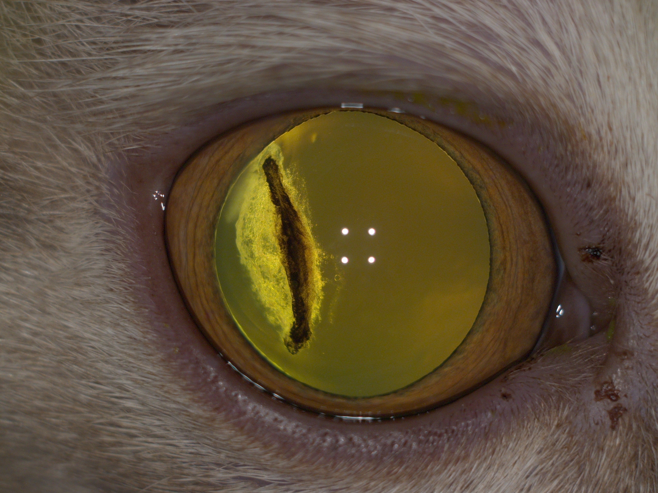
EYE SURFACE IMAGING
Document eye surface conditions
Obtain eye surface images with the anterior module of the Optomed Aurora IQ. Monitor corneal abrasions and ulcers with fluorescein staining using the cobalt blue filter on the anterior segment module.
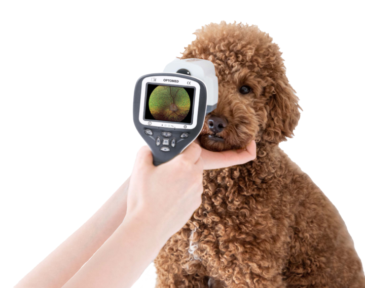

Portable and affordable retinal imaging
The Smartscope PRO is a small-sized handheld fundus camera with a 40 ° field of view and non-mydriatic operation. Examination possibilities can be further expanded with an anterior module that includes white LEDs for color imaging and blue LEDs for fluorescein imaging.
.
CLINICAL EVIDENCE
Optomed Fundus Cameras in Veterinary Research
Optomed handheld fundus cameras have been used to image several animal species* in more than 15 peer-reviewed scientific publications.
Fundus imaging in 208 cats
In a 2025 study, Optomed Aurora was used to capture fundus images from 208 cats (416 eyes) without pupil dilation. The researchers reported high image quality for centration, clarity and visible retinal area.
*Optomed cameras have been used for retinal imaging in cat, dog, cattle, ring-tailed lemur, macaque, fallow deer.
Read the study here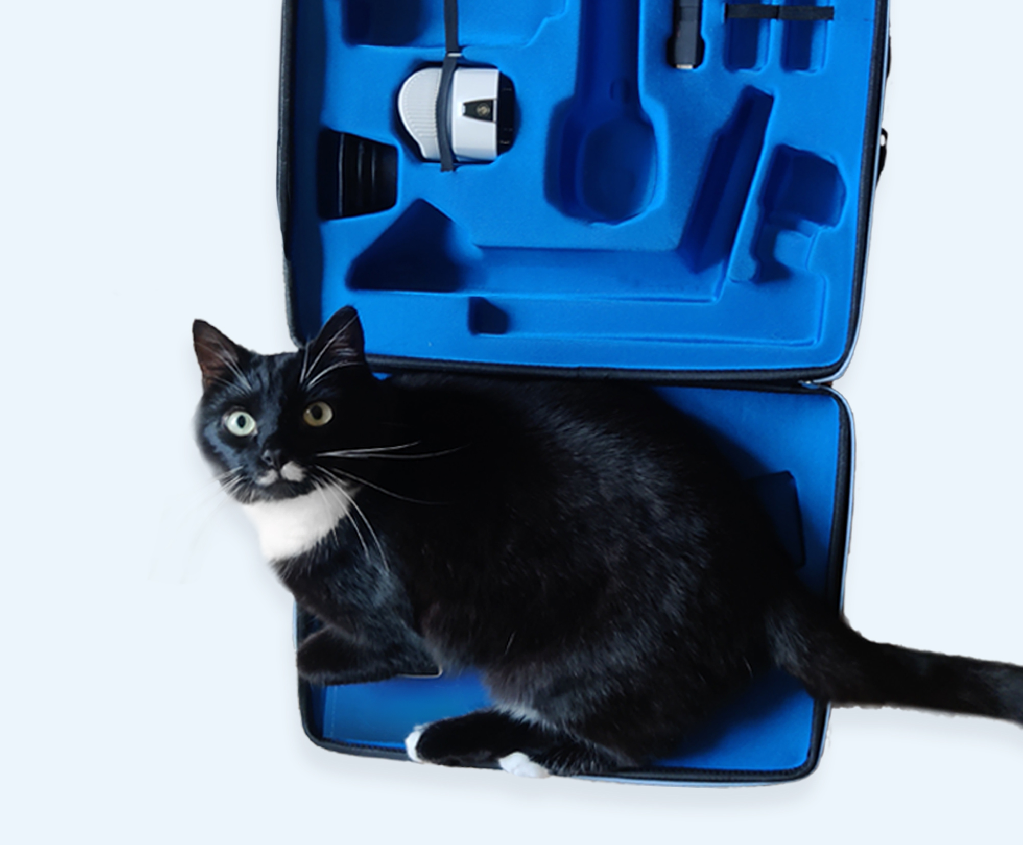
Read what our users think about us
A retinal image allows a more detailed view of findings for a later review than a written description
Kaisa Wickström
Certified ECVO Panelist
Licentiate of Veterinary Medicine
Veterinary clinic Kamu,
Oulu, Finland
I use the Optomed Aurora IQ camera for the documentation and follow-up of retinal findings. A retinal image allows a more detailed view of findings for a later review than a written description. The camera has also been used in imaging retinal conditions suspected to be genetic. In these cases, it has been possible to send the images directly to a canine genetic research group to be used in scientific studies.
Features that I like in Optomed Aurora IQ include a wide field of view and an almost silent operation; a loud sound for example during focusing can frighten dogs and could prevent image capture. Infrared aiming light of the camera eases the imaging a lot. It is possible to capture high quality images even though the pupil is not dilated. At first, I had doubts about the length of the camera, but image capture from even active dogs has succeeded well after practicing the technique. The camera is practical for the follow-up of diseases and particularly useful in demonstrating changes in the animal’s eye to the customer directly from the camera display.
Optomed Aurora has been a great tool for us to use in a wide variety of species, and students love being able to see lesions
Dr. Ellison Bentley
University of Wisconsin-Madison
Optomed Aurora fundus camera is easy to use in veterinary ophthalmology and provides excellent quality images
Dr. Carrie Breaux
Mountain West Veterinary Ophthalmology
Contact us