Central retinal artery occlusion (CRAO) is devastating and the incidence is estimated to be one in 100,000 people. Unfortunately, CRAO is a condition that usually presents with sudden, painless monocular vision loss. This often means medical personnel can do little to help.
Even more challenging is that the processes available for diagnosing CRAO are not well-suited for emergency response. They take up precious minutes, during which the patient’s sight can degrade rapidly.
The good news is that new technology is giving emergency responders a tool to help speed their recognition and diagnosis. It has the potential to make dramatic improvements in retinal artery occlusion fundoscopy, leading to faster treatment.
It’s also about more than just vision. Retinal artery occlusion can often be a precursor to cerebral stroke, and improved retinal artery occlusion fundoscopy can help doctors initiate life-saving treatments sooner. Retinal cameras can streamline the assessment of CRAO.
Recognizing and Treating Central Retinal Artery Occlusion
When a patient arrives in the ER indicating that they have lost sight in one eye, it likely indicates CRAO, so it’s an emergency and the patient needs immediate attention.
The problem is that current diagnostic systems are not fast. They take up time that the patient doesn’t have and present several obstacles to clearly identifying the location and type of occlusion.
For instance, the use of ophthalmoscopes to visualize the retina in many cases requires dilation of the patient’s pupils. Dilating drops can take a half hour or more to begin to work, which does not help with a patient’s prognosis.
Other diagnostic methods such as OCT, fundus fluorescein angiography, MRI, or CT scan can show profound imagery to help with diagnosis.
Simplifying Retinal Artery Occlusion Fundoscopy in the ER
Mobile fundus cameras won’t replace those more sophisticated methods, but they do provide a faster pathway to information that ER doctors and their staff can use to take action more immediately.
One distinguishing characteristic of CRAO diagnosis is that it requires the ability to recognize its clinical features, such as a cherry-red spot, retinal opacity, optic disc pallor, and optical atrophy, among others.
Mobile fundus cameras capture still photos, time-lapse photos, and video that can help ER doctors spot those signals more rapidly and with more precision.
The cameras are non-invasive. They are lightweight and handheld and can be operated by a single staff member or technician. Doctors and staff can easily carry the device from room to room.
Even more importantly, the cameras capture images with a wider field of view, enabling doctors to examine more of the eye more quickly for problem spots.
The cameras are also non-mydriatic, meaning they can capture images without having to wait for pupil dilation drops to work. The key is getting as wide a view of the retina as possible in the shortest amount of time.
Finally, mobile fundus cameras can help speed communication with specialists by linking to hospital systems. This is particularly important in rural areas, which may not have ready access to on-site specialist care for what is generally a rare event.
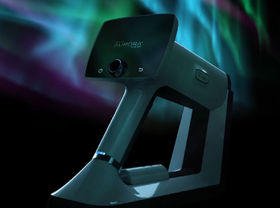
Saving Sight with the Optomed Aurora
Optomed is a leading manufacturer of handheld fundus cameras globally. The team is committed to innovating eye care to reduce unnecessary blindness.
The company’s Aurora camera system is a mobile handheld retinal camera that is ideal for use in the hands of primary care and emergency room physicians, as well as their respective staff members.
The Aurora is easy to learn to use. It can be implemented immediately upon a patient presenting with sudden blindness to capture clear images and videos that give a view into the health of a patient’s vasculature.
The imagery captured in the ER becomes part of the patient’s record and is immediately shareable over PACS networks.
CRAO is a medical emergency that can be frightening to patients and their families and is hard on physicians and their staff. Retinal cameras can improve retinal artery occlusion fundoscopy and take away some of those burdens.
Contact Optomed to Help
Working together with primary care and emergency room personnel, Optomed aims to provide technology for the improved diagnosis and treatment of eye diseases, such as diabetic retinopathy and other conditions, with the goal of minimizing blindness.
Patients experiencing CRAO need IV tPA within 4.5 hours for the best outcomes. That means you need to act FAST. Our mobile fundus cameras are easy to use for staff, enabling teams to easily confirm or refute CRAO in the ED. Digital image sharing helps emergency teams save critical time by streamlining communication.
Want to learn more about how we’re enabling neurologists, neuro-ophthalmologists, and emergency staff to confirm or refute CRAO faster and easier than ever?
Share this insight