The examination and documentation of retinal health are critical components of every complete physical examination. Conducting an accurate and thorough eye examination is critical to the diagnoses of several sight- and life-threatening medical conditions. The Association of American Medical Colleges, the American Academy of Ophthalmology, and the ICO emphasize that medical students, at minimum, should be able to perform a basic eye examination with fundoscopy and describe their observations proficiently.
Traditionally, ophthalmoscopes have been used to visualize and document the retina. The ophthalmoscope has been a valuable diagnostic tool for general physicians and eye care professionals for over 150 years. However, as technology has advanced, its limitations have become increasingly apparent. In this article, we will explore the drawbacks of the ophthalmoscope and make the case for upgrading to a more advanced and accurate diagnostic tool – the non-mydriatic handheld fundus camera.
Drawbacks of the Ophthalmoscope
One of the main drawbacks of the ophthalmoscope is its steep learning curve. Many professionals note that using the ophthalmoscope requires significant skill and training, which can make it challenging for healthcare professionals who do not specialize in eye care. However, in medical education, it is not easy to incorporate such specific training in the curriculum or to objectively assess the required operational skills. Add in the fact that dedicated ophthalmology curriculum hours and clinical teaching has declined, and ophthalmology rotations are only required in less than a fifth of medical schools and you can see why many medical school graduates feel they are inadequately trained and lack confidence in their ability to perform this important clinical skill upon entering residency training.
Only about 30% of second year medical students could identify the optic nerve of a patient when performing an eye exam using a direct ophthalmoscope. The Association of University Professors of Ophthalmology (AUPO) notes in a recent survey that most program directors believe that only about half of their students meet the AUPO standard for identifying and dealing with eye problems; even after completing medical school and residency.
A popular study found that only 14% of patients presenting to the emergency department at a large academic center with symptoms or conditions warranting ocular fundus examination had ophthalmoscopy performed by the physician. This suggests that trained physicians are hesitant to perform the test, despite the medical necessity to do so.
Furthermore, the ophthalmoscope provides a limited view of the eye, making it difficult to assess the retina and optic nerve in detail. With a viewing field of 5 to 10 degrees and a 15× magnification, the direct ophthalmoscope makes it challenging to obtain a proper visualization of the optic nerve head, not to mention regions away from the nerve. The use of an ophthalmoscope also requires a high level of cooperation and compliance from the patient. Patients must remain still , which can be challenging, especially for young children or patients with neurological or cognitive impairments. The fleeting view obtained may be small, blurry, or difficult to interpret, making it challenging to identify subtle changes or abnormalities in the retina. This can result in inaccurate diagnoses and unnecessary referrals, which can be costly and time-consuming for patients and healthcare providers alike.
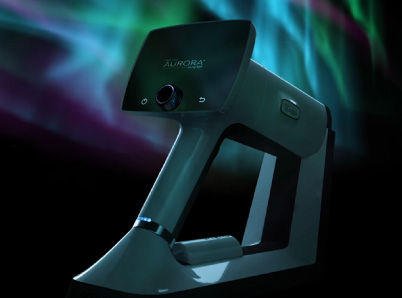
The Case for Fundus Cameras
Handheld fundus cameras are compact, portable devices that can capture high-quality images of the retina. They offer several advantages over ophthalmoscopes, including higher image quality, increased efficiency, and the ability to document changes in the retina over time. These benefits can lead to more accurate diagnosis and personalized treatment plans for patients, which may ultimately help prevent irreversible damage to the retina.
These cameras capture wider-field, high-resolution images of the retina and optic nerve, providing a more detailed and accurate assessment of the eye’s health. In fact, studies have shown that fundus cameras are significantly more accurate than ophthalmoscopes when it comes to diagnosing eye conditions such as diabetic retinopathy and glaucoma.
In one study emergency physicians used non-mydriatic fundus photographs more frequently than they performed direct ophthalmoscopy, and their detection of relevant abnormalities improved. Ocular fundus photography often assisted ED care even when normal. Students preferred fundus photography for examining the fundus, with 70% of medical students finding it easier and less frustrating than using ophthalmoscopy.
In addition, fundus cameras are more patient-friendly than ophthalmoscopes. Patients often find the dilation, constant bright light, and close proximity of the ophthalmoscope uncomfortable, which can make it difficult to obtain accurate readings. Fundus cameras, on the other hand, are non-invasive and require no direct contact with the eye, making them a more comfortable option for patients.
Handheld fundus cameras are also versatile, as they can be used in a variety of settings, from remote areas with limited access to specialized medical equipment to emergency departments where quick assessments of retinal health are necessary.
Fundus photographs taken by non-ophthalmology practitioners and non-physicians alike consistently show the ability to obtain high quality photographs in greater than 90% of patients when using fundus cameras. They show no significant differences in quality whether the images were taken by a physician or a technician. Fundus photos are the gold standard when diagnosing diabetic retinopathy and non-mydriatic handheld fundus cameras such as the Optomed Aurora IQ show high sensitivity and comparable performance to bulky desktop machines when diagnosing from fundus images.
Conclusion
While the ophthalmoscope has been a valuable diagnostic tool for many years, it is clear that it has its limitations. There is evidence that direct ophthalmology, even by ophthalmologists, is sub-optimal in detecting common abnormalities versus fundus photographs. Students even learn better when using fundus photographs. As technology continues to advance, healthcare professionals should consider upgrading to more advanced and accurate diagnostic tools such as non-mydriatic handheld fundus cameras like the Aurora IQ.
Share this insight