When it comes to diagnosing and treating eye diseases in animals, accurate and efficient diagnosis is key. However, traditional diagnostic methods for veterinary ophthalmology, such as direct ophthalmoscopy, can be challenging, time-consuming, and require specialized training. As the field of veterinary medicine continues to evolve, new technologies and techniques are emerging to provide enhanced diagnostics and care for animals. That’s where handheld fundus cameras come in – these portable and user-friendly devices are transforming the field of veterinary ophthalmology and providing more accurate and efficient diagnosis and treatment options for animals.
In this blog post, we will explore the benefits of using fundus cameras in veterinary ophthalmology, including sharper insights, timely diagnosis, faster workflows, and improved patient comfort. We will also delve into the essential role that fundus cameras play in advancing veterinary ophthalmology and how they are helping veterinarians capture clarity and precision in retinal examinations.
Overview of Handheld Fundus Cameras
Handheld fundus cameras are specialized cameras that can capture detailed images of the retina, the light-sensitive tissue at the back of the eye. These cameras typically use LED illumination and a high-resolution image sensor, while providing efficiency and portability.
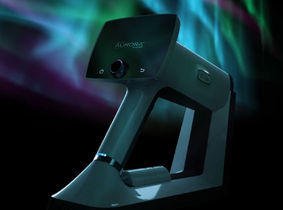
The use of handheld fundus cameras is becoming increasingly common in veterinary medicine for the diagnosis and management of ocular diseases in animals of many different species. Some like the Optomed Aurora IQ can also take high quality pictures of the front of the eye and its surrounding structures.
“The Optomed Aurora has been a great tool for us to use in a wide variety of species, and students love being able to see lesions.” Ellison Bentley, University of Wisconsin-Madison
Advantages of Handheld Fundus Cameras
- Portability: Handheld fundus cameras are portable and easy to use, making them ideal for use in the field or in veterinary practice. They can move to the exam room or barn where the patient is, or in between clinics as needed, increasing your workflow efficiency.
- Affordability: Handheld fundus cameras are generally more affordable than confocal scanning laser ophthalmoscopy devices, making them a more accessible option for small animal practices or veterinary clinics with limited budgets.
- Ease of use: Handheld fundus cameras are designed to be easy to use, with intuitive controls and a lightweight, ergonomic design that minimizes operator fatigue. You are in control of how the camera is positioned and when to take the image of your non compliant animal patient.
- Patient comfort: The handheld design of these cameras allows for quick and comfortable imaging of the retina without the need for a general anesthesia or tranquilization, which can reduce stress on the animal and minimize the risk of complications. Their nonmydriatic design can work without dilation in some cases as well.
- Adaptability: Some handheld fundus cameras like the Optomed Aurora IQ can also take pictures of the front surfaces of the eye. A cobalt blue light filter can also be utilized for hyperfluorescence, enabling documentation and management of corneal problems.
- Quick diagnosis: Immediate on camera viewing of these high-quality photographs can lead to faster workflows within the clinic. When ophthalmic emergencies happen, time is of the essence to save vision. Infrared digital photography can increase the contrast of structures, monitoring of pigment changes, and increase visualization through cloudy corneas.
“The Aurora fundus camera is easy to use in veterinary ophthalmology and provides excellent quality images.” Dr. Carrie Breaux, Mountain West Veterinary Ophthalmology
Applications in Veterinary Medicine
Handheld fundus cameras have a wide range of applications in veterinary medicine. Some of the most common uses include:
- Diagnosis and management of systemic and ocular diseases: Handheld fundus cameras allow veterinarians to quickly and accurately diagnose ocular diseases such as retinal detachment, glaucoma, and uveitis.
- Herd based ophthalmic screening: Getting field-based livestock transported to a hospital for screening is difficult, time consuming, and expensive. Using a portable, nonmydriatic fundus camera has been shown to be an effective and easy solution.
- Screening for genetic disorders: Some breeds of animals are predisposed to certain genetic disorders that can be detected through a fundus examination. Handheld fundus cameras make it easier to screen for these disorders in the field or in a clinical setting.
- Monitoring disease progression: Handheld fundus cameras can be used to monitor the progression of ocular diseases over time, which can help veterinarians adjust treatment plans as needed.
- Teaching and research: Handheld fundus cameras are also useful tools for teaching veterinary students and for conducting research on ocular diseases in animals. Showing examples of retina diseases and eye structures to students who may have difficulty visualizing them with an ophthalmoscope.
Conclusion
Handheld fundus cameras are becoming increasingly popular in veterinary medicine due to their portability, affordability, and ease of use. With continued advancements in technology, handheld fundus cameras have become higher quality and of greater clinical importance.
Handheld fundus cameras such as the Optomed Aurora IQ have been used on many species of animals including rodents, felines, canines, equines, bovines, porcines, birds, and zoo animals of many shapes and sizes.
Share this insight