Fundus photographs have emerged as a valuable tool in evaluating cardiovascular and neurologic risk. Robust data from clinical and population-based studies consistently show significant associations between elevated blood pressure and retinal changes. Identifying new hypertensive retinal findings is strongly linked to elevated blood pressure levels. This comprehensive guide explores the profound connection between chronic and acute retinal and cerebral vascular diseases, with specific features like retinopathy, retinal artery occlusion, and increased retinal vein caliber predicting future cerebrovascular events. Discover how utilizing retinal vasculature findings can help assess and stratify individuals based on their risk of cerebral vascular disease. By leveraging fundus cameras, physicians and practice managers can ensure thorough eye evaluations, early detection of retinal microvascular abnormalities, and timely interventions to enhance patient outcomes and overall well-being.
Retinal findings can be categorized into two groups. The first involves the retinal arterioles directly, including generalized and focal arteriolar narrowing, and arteriovenous nicking. These changes act as markers of cumulative, long-term hypertension-induced damage and have been independently correlated with blood pressure levels measured 5-8 years before the retinal assessment. On the other hand, the second group encompasses changes in the retina itself, such as hemorrhages, cotton wool spots, microaneurysms, macular edema, and exudates. Focal arteriolar narrowing, retinal hemorrhages, microaneurysms, and cotton-wool spots are indicative of acute changes in blood pressure and are associated with concurrent blood pressure measurements.
The clinical significance of these retinal findings is profound. Specifically, retinopathic changes, but not vasculopathic ones, have been linked to double the risk of incident congestive heart failure, even among individuals with otherwise low cardiovascular risk. Moreover, the presence of hypertensive retinopathy doubles the risk of left ventricular hypertrophy and elevates the risks of renal dysfunction and cardiovascular mortality. Notably, sub-analyses of the Beaver Dam Eye Study reveal that individuals exhibiting retinal microaneurysms and retinal hemorrhages are twice as likely to experience cardiovascular events leading to mortality compared to those without these signs.
Furthermore, compelling evidence indicates an increased risk of ischemic heart disease mortality associated with arteriolar narrowing and decreased retinal arteriolar tortuosity. These findings underscore the crucial role of retinal examination in identifying patients at risk of cardiovascular disease and emphasize the importance of integrating it into routine comprehensive risk assessments. By leveraging retinal microvascular examination, physicians can detect early markers of cardiovascular risk and tailor preventive interventions to enhance patient outcomes.
In the United States, a stroke occurs every 40 seconds, and it is the primary cause of long-term disability. In addition to clinical assessment, the primary diagnostic modality to assess stroke is non-contrast head computed tomography (CT); additional modalities might be acquired for further assessment. Retinal disease changes can be a harbinger of cerebral microvascular changes – and thus brain pathologies, with fundus photography being a non-invasive key in this screening process.
Fundus images could also be used to analyze the retinal age gap and its association as a predictor of stroke risk. One study analyzed fundus images of a population that did not have a history of stroke and followed them for 5 years. Results indicated that with each extra year in the retinal age gap, the risk of stroke went up by 4%. And those with a larger retinal age gap in the fifth quartile had a much higher risk of stroke. Furthermore, looking at the retinal age alone by analyzing their fundus photos was comparable to using the well-established risk factor-based model. In patients with diabetes mellitus and hypertension, patients with vascular retinopathy with vessel rarefaction had a higher risk for recurrent stroke. Retinal artery or retinal vein occlusions and stroke represent acute manifestations of vascular disease affecting both the retina and the brain. Simultaneous occurrences of blood clots in both retinal and brain circulations are frequently observed.
Another study analyzing blood pressure and its risks with retinal vascular events found that elevated blood pressure, stage 1 hypertension, and stage 2 hypertension were all associated with higher retinal vascular occlusion risks than normal blood pressure. Controlling hypertension appears to reduce the risk of subsequent retinal vascular occlusion; however, the incidence rate was still significantly higher than that in persons who maintained a normal blood pressure. The retinal circulation serves as a potential marker of cerebral vascular disease, primarily due to its shared origin and drainage with the intracranial circulation. Furthermore, its direct visualization using funduscopy makes it a valuable tool for assessing and monitoring related conditions.
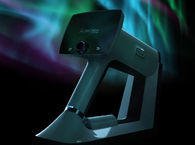
Fundus photography offers a more detailed and comprehensive view of the retina and optic nerve, aiding in early detection and precise diagnoses of retinal microvascular abnormalities and their association with cardiovascular and neurologic disease risk. The important retinal structures and minor retinal changes can be easily visualized in fundus images and these can be sent for consultation to a colleague via telemedicine. Optomed is committed to making improved screening available to as many patients as possible through affordable, intuitive, portable cameras that produce world-class images.
Share this insight