Advances in medical imaging technology are transforming the early detection of eye disease by providing retina scan capabilities that produce detailed views.
Using new digital cameras designed specifically to examine the eye, doctors are now better able to spot signs of conditions like glaucoma, diabetic retinopathy (DR), and age-related macular degeneration.
Lightweight and portable, these new cameras enable fast and efficient eye exams that allow the retina scan to become part of the regular workflows of today’s busy primary care offices.
In providing primary care physicians (PCPs) with this new capability, these cameras are increasing the chances for medical interventions that can prevent these conditions from progressing to blindness.
Digital retina cameras also may help PCPs diversify their practices by expanding the support that they offer patients in a sustainable, cost-effective way.
Systematic Screenings Save Eyesight
DR, glaucoma, and age-related macular degeneration are conditions that develop slowly. By the time that they are detected, it is often too late.
The current model for detecting these conditions is built around the ophthalmoscope, a century-old medical device. Its use at primary care offices has declined because some say that it is too hard to use and the images are difficult to interpret. As a result, early detection depends on patient compliance with referrals to specialists, which are difficult to ensure.
Studies have shown that early and regular retina scans can make an enormous difference in preventing eye diseases from progressing to blindness. For example, the incidence of sight-threatening DR in England fell dramatically after the country introduced annual diabetic eye screenings.
Similarly, in Iceland, researchers found a lower prevalence of DR following the implementation of a national diabetic retina scan program in that country. Researchers found that the screening programs had the potential to significantly “decrease the likelihood of blindness” in diabetic patients. The advent of modern imaging technology may well build on those results, helping save the sight of millions around the world.
Benefits for Physician Practices
The ability to do a preventative retina scan has become more compelling with the development of retinal cameras that can identify the telltale signs of sight-threatening eye disease.
The biggest benefit of the cameras is that they are non-mydriatic: they do not require a patient’s eyes to be dilated to produce effective imagery for detection. This contrasts with the ophthalmoscope. Pupil dilation can take up to a half hour to take effect, which is ineffective for office workflow and inconvenient for the patient.
Retinal cameras are also handheld, making them easy to move from room to room or to be included in kits for visits to clinics. They also offer a wider field of view, making the retina scan easier for doctors.
Some camera systems for the eye can be integrated with artificial intelligence software that further streamlines the retina exam. This software adds decision-making support by rapidly identifying problem areas at an accuracy rate that is equal to or better than manual image reviews.
How Optomed Aurora Improves Workflow and Patient Care
Optomed has long recognized the benefits of early eye screenings and has become one of the world’s leading innovators in developing tools that allow for a more efficient retina scan. The company’s goal is to make eye screenings available for everyone, wherever they are.
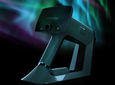
The Optomed Aurora Handheld Fundus Camera provides next-level imaging capabilities for the examination and documentation of the retina and anterior of the eye for various eye diseases and neurological disorders. The camera can produce both still images and video of the retina scan for review. It comes with integrated image quality analysis to guide first-time users toward success in capturing images.
Offering a 50-degree-wide field of view, the Optomed Aurora gives doctors a handheld device designed for ease of use. The camera relies on the use of infrared light to target the fundus and has autofocus, auto exposure, and high-contrast modes to pinpoint areas that need closer examination.
Significantly, the Aurora system is easily networked, able to connect to patient health records or hospital networks to provide documentation for the future. By retaining previous images that can quickly be retrieved, doctors can study changes over time and explore if warning signs were missed.
Optomed fundus cameras also come with optional cloud connectivity to AI tools, which can help pinpoint areas that need further review.
The Case for Optomed
Advanced imaging technology is poised to transform the prevention and treatment of devastating eye disease. With digital retinal cameras such as the Optomed Aurora, doctors can now build routine eye exams into their everyday practice. The system enables you to manage the needs of your patients and the flow of activity in your practice. The tools are within our grasp to help prevent unnecessary vision loss from progressive eye diseases.
Here at Optomed, our mission is to help save the vision of millions of people. By integrating our software and artificial intelligence solutions with our camera, we enable eye screening for everyone, wherever they are. To see how we can equip you to save the sight of more patients, schedule a free consultation today!
Share this insight