The frightening prospect of losing the ability to see is real for an estimated 93 million adults across the United States. The CDC also reported that unfortunately, only about half of people at risk visited an eye doctor for a screening for retina problems in the past year.
People are facing severe vision impairment and blindness due to glaucoma, diabetic retinopathy, age-related macular degeneration, and other eye diseases that interfere with the proper functioning of the retina. The widespread nature of the problem highlights the urgency of early detection. The sooner that retina problems can be spotted, the greater the chance of stopping the progression of the disease.
Early detection of retina problems can be dramatically improved. Today, it relies largely on the direct ophthalmoscope, a device invented more than 150 years ago. Many in health care see the ophthalmoscope as inefficient and hard to use, especially for non-eye care specialists. The device also has several limiting factors that make early detection a challenge.
Driven by the digital age, new tools are now being developed that can help doctors, especially primary care doctors, screen more patients in less time, thereby helping to reverse the threat posed to eyesight from retina problems. With these tools, doctors in primary care settings have a powerful opportunity to help patients avert blindness.
The Challenge of Detecting Retina Problems
Glaucoma, diabetic retinopathy, age-related macular degeneration, and eye diseases tend to be asymptomatic. They cause tiny, undetectable changes until the damage is irreversible.
Despite the best efforts of primary care doctors to encourage patients to seek preventive eye exams even when they feel adherence lags. Health care has long needed the tools and processes to make it easier for more doctors to screen more patients in less time.
For eye screenings, doctors and specialists primarily use the direct ophthalmoscope, which has several limiting factors that make the mastery of it “a dying art,” according to some researchers.
In general, ophthalmoscopes can be hard to use, especially without the patient having their pupils dilated. The process of pupil dilation takes up to a half-hour to take effect. The ophthalmoscope also requires practice in order to get the techniques down to make reliable assessments. Finally, ophthalmoscopes also offer a narrow field of view of 5 degrees or less.
New Ways for Doctors to Help Avert Blindness
The good news is that technology is being developed that offers a better way. Specifically, fundus photography is a promising alternative to the ophthalmoscope.
Fundus photography involves the use of digital cameras that allow doctors to see a wider portion of the retina. They can produce still and video images of the back of the eye that can be archived, studied offline, and shared easily with specialists.
Studies show that fundus photography can significantly increase the ability of health care professionals to properly diagnose retina problems, such as diabetic retinopathy. The next level in the technology involves the use of smartphones with special attachments to capture imagery.
Fundus photography and machine learning/artificial intelligence and other software capabilities offer many advantages for detecting retina problems compared to the traditional ophthalmoscope.
One of the most significant is that retinal camera systems are easier and less expensive to use, making them available to a broader cross-section of medical professionals.
Retinal cameras are also touchless and mobile. Doctors can take them on home visits or to rural clinics to do proper eye screenings.
Last but not least, the digital cameras are network connected. Images can be transmitted via PACS or EHR systems for quick evaluation by ophthalmologists and retained for future comparison to judge changes over time.
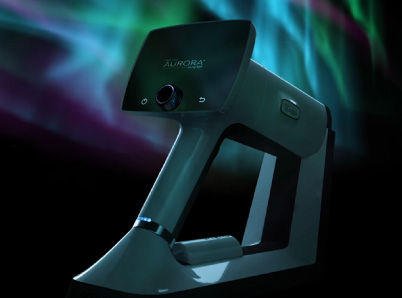
Optomed Delivers Early-Detection Solutions
For retinal cameras to be successful in addressing early detection, they must be easy to use for everyone, cost-effective, efficient, and easily connected to medical networks. Optomed meets those needs on every front.
The Optomed Aurora is a non-mydriatic system, meaning that it can capture images without patients having their pupils dilated. The camera has been designed to enable easy examination and documentation of the retina and anterior of the eye for various eye diseases and neurological disorders.
The Aurora has a wide, 50-degree field-of-view to allow for a more detailed review. The camera also includes a high-contrast optical design that helps detect small early-phase retinal changes.
The camera is lightweight, handheld, and portable. Doctors can take it to patients, wherever they are, to conduct screenings for eye disease.
The medical evidence is clear: the earlier that doctors can detect retina problems, the more likely that patients can receive sight-saving treatments, including new techniques that are showing great promise.
Primary care doctors are poised to be an essential part of that solution, helping spot signs of eye disease more reliably and cost-effectively than ever.
Here at Optomed, our mission is to help save the vision of millions of people.To see how we can equip you to save the sight of more patients, schedule a free consultation today!
Share this insight