Age-related macular degeneration is a disease that PCPs tend to associate with aging. The Beaver Dam Eye Study found that pure geographic atrophy (late age-related macular degeneration) was 16.6 times more likely to occur in patients who were over seventy-five than in those who were under seventy-five at the baseline. But when the Third National Health and Nutrition Examination Survey examined fundus photographs of 4,007 US patients, the estimated prevalence of the disease was 9.2% among non-institutionalized civilian persons forty years of age and older. But age-related macular degeneration appears before age sixty in Black and Hispanic patients, who are underserved. Age-related macular degeneration was detected at 53-69% higher rates for Black and Hispanic patients than for white patients before the age of sixty and then at 50-63% higher rates for white patients after the age of sixty.
Black and Hispanic patients in the US also have lower rates of annual eye exams and higher rates of diabetes, so in addition to age-related macular degeneration, they are also at risk of diabetic retinopathy. The implication of these findings is they need to get eye exams in their forties and fifties and not wait until they are in their sixties, as advanced, exudative disease may have already developed.
Primary care providers have an important role in timely referrals for the treatment of age-related macular degeneration and diabetic retinopathy. But scalable inclusion of screenings for these conditions is not possible with traditional diagnostic tools.
The utility of the direct ophthalmoscope in detecting age-related macular degeneration is nil.
In the twentieth century, many physicians were taught direct ophthalmoscopy early in their training. It is a familiar instrument, with some uses in clinical exams.
Ophthalmoscopes are available and affordable, and they can be carried in the doctor’s coat pocket. They provide a 15x magnified view of the posterior pole, facilitating an appreciation of small, dynamic changes of the ocular fundus. They can be used in clinical settings where other forms of ophthalmoscopy are not available.
But the direct ophthalmoscope is inadequate for screenings for age-related macular degeneration and other maladies of aging eyes.
The most significant limitation of the direct ophthalmoscope is the narrow field of view. In an emmetropic patient, the direct ophthalmoscope permits examination of an area of about two disc diameters, or 7 mm2, of retina simultaneously in focus. The field of view is smaller in a myopic patient.
Given that the surface area of the average human retina is approximately 1,200 mm2, the physician would need to make at least 170 systematic observations of the retina to avoid missing even a fairly large lesion that was two disc diameters in size.
The classic direct ophthalmoscope’s five-degree field of view is too small to resolve all of the relevant details of the optic disc and retina at once. An enhanced ophthalmoscope offers a twenty-five-degree field of view but at a lower magnification. Pharmacologic pupillary dilation compensates for these deficiencies but takes thirty minutes or longer and is objectionable to patients.
The confluence of these factors means that even in the hands of the most skilled practitioner, it is not possible to visualize the retina with a direct ophthalmoscope. There is considerable opinion among authorities in academic medicine that the technique should not even be taught to physicians. However, modern technology brings screening for age-related macular degeneration back to the primary care physician.
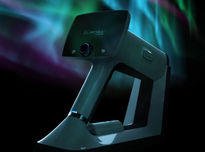
Timely diagnosis is necessary for timely care and is possible with Optomed.
- Optomed Aurora facilitates non-mydriatic exams.
- Optomed Aurora has multiple applications in primary care, not just age-related macular degeneration, diabetic retinopathy, and other conditions common in aging eyes.
- Portability makes Aurora ideal for pediatric care, geriatrics, homecare, and screening camps.
- CMS reimbursement guidelines suggest that Optomed may enhance practice revenues.
A documented complete macular examination is necessarily prerequisite to determining the presence and severity of age-related macular degeneration, so a decision can be made as to the benefits of ongoing supportive therapies, such as antioxidant vitamins, or a referral to early intervention, such as laser photocoagulation surgery. Periodic re-assessment is necessary to determine whether there is progression of the disease and to plan the on-going treatment.
There is no data on the frequency of reexamination for age-related macular degeneration in primary care, but parallel data for key assessments for glaucoma, cataracts, and diabetic retinopathy suggest that significant gaps in examination are likely. To encourage ongoing monitoring for age-related macular degeneration, CMS has created codes for billing screening exams.
Optomed potentiates profitable and ethical ancillary care in primary practice.
There is general agreement that professionals and patients alike tend to equate new technology with a higher standard of care. However, capital investments in some new technologies create financial pressures on practitioners to use those devices in routine examinations.
This is not an issue with Optomed. The Optomed Aurora technology is affordable and scalable, and used in appropriate patient populations, it generates a necessary and ethical benefit to patients. Ancillary procedures with a medical indication, such as screening for age-related macular degeneration, meet requirements for reimbursement by insurance and CMS.
Here at Optomed, our mission is to help save the vision of millions of people. By integrating our software and artificial intelligence solutions with our camera, we enable eye screening for everyone, wherever they are. To see how we can equip you to save the sight of more patients.
Share this insight