One of the biggest health problems in the United States today is diabetes. It’s estimated that 37 million people have diabetes. Of those, 95% have type 2 diabetes. Diabetic retinopathy is one of the most common causes of blindness and can begin anywhere from three to seven years before a type 2 diabetes diagnosis. The typical way to screen for diabetic retinopathy is through a fundus screening. A specialized camera takes a picture of the rear of the eye, called the fundus. It’s a straightforward process, but many patients don’t know that diabetes can affect the eyes, and primary care physicians might not be up to date on screening methods.
Even if both the patient and the physician are aware of the potential problem, access to fundus screenings is limited in most places, so when the patient finally gets screened, there is already irreversible damage. Almost 50% of patients in the US are not screened. Even in the UK, where screenings are free, it’s estimated a third of patients are not screened in time.
Technology has produced much more reliable screening methods, but they can’t be used without a reliable digital framework to build upon. Also, fundus cameras are often large, bulky instruments that are fixed in one location, most often in ophthalmologist clinics. Many people do not live near such a clinic or otherwise have the ability to visit one with or without a referral. Further complicating the matter is that insurance has not covered it until recently; it was only in 2018 that Medicaid/Medicare even had a category for retinopathy screenings. Patients covered by either usually have limited funds and cannot pay for the process out of pocket.
If patients are screened on a reliable basis, time and money are saved. Fundus screenings save money on treatment by catching retinopathy early, before it may cause symptoms, and time spent treating it can be reduced. The best way of doing so is to make fundus screenings more readily available in easier-to-access places. If a screening is not tied to certain hospitals and can be done in an ordinary doctor’s office or local clinic, patients can be screened without the hassle.
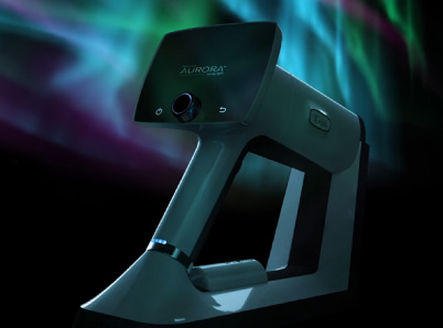
A handheld fundus camera can easily accomplish this. Doctors can perform screenings wherever they’re needed, even in extremely remote locations. Unlike older models, where the images have to be analyzed by someone trained in reading them, these cameras can be connected to an artificial intelligence database that can assess the data for them. The database can even schedule appointments and send reminders of follow-ups to patients who have already been screened. The only thing that needs to be done is to train the operator in the use of the camera itself.
A fundus screening, while primarily for diabetics, can also be used for other conditions. Since many other conditions can affect the minute blood vessels of the eye, the ability to see them without an invasive procedure could potentially lead to the diagnoses of other conditions. Glaucoma can be detected from early fundus photography because it can reveal early pressure on the optic nerve. Narrow blood vessels in the fundus area can indicate damage from high blood pressure. Dark spots in the fundus might be an early sign of melanoma. The same advantages that portable fundus screenings bring to eye exams also apply to any other condition that affects the blood vessels of the eye.
While access to the fundus screening cameras themselves, as well as training for how to use them, still needs to be in place before any widespread screenings can begin, their potential availability is a big step toward solving the screening problem, and that is a good start.
Here at Optomed, our mission is to help save the vision of millions of people. By integrating our software and artificial intelligence solutions with our camera, we enable eye screening for everyone, wherever they are. To see how we can equip you to save the sight of more patients, schedule a free consultation today!
Share this insight