Early detection offers the best defense against the effects of progressive, blinding eye diseases such as glaucoma, diabetic retinopathy, and age-related macular degeneration.
Successful early detection relies on the ability of doctors to gain a clear view of the fundus. With the proper tools, providers can spot lesions and other signs of progression, evaluate them quickly, and provide follow-up care or specialist referrals.
Currently, outdated diagnostic technology—specifically, the ophthalmoscope—has obstacles to earlier detection. Many view the ophthalmoscope as hard to use and disruptive to workflows in emergency rooms and primary care offices.
But spurred by innovations from companies such as Optomed, the healthcare system is overcoming those obstacles. New imaging systems are making it easier for doctors to take a critical look at the eye’s fundus and provide warning that allows for early action to prevent sight loss or blindness.
These new tools are maturing at the right time, as researchers are forecasting a surge in diabetes worldwide that could lead to a significant increase in cases of diabetic retinopathy.
Importance of Visualizing the Retina
Diabetic retinopathy, age-related macular degeneration, and glaucoma are progressive conditions that make small changes slowly over years. By the time that problems emerge, it’s often too late.
These diseases are vascular in nature. The fundus is the only place in the body where doctors can directly observe micro-circulation to identify oncoming problems with eyesight.
Current technology does well in helping spot the presence or absence of telltale retinal lesions indicative of disease. But the approach falls short in providing simple and efficient ways of identifying those at the highest risk of progressing to blindness.
The need for new capabilities to evaluate the seriousness of progressive eye disease is urgent. Diabetes is an international epidemic, expected to reach more than 640 million adults by 2040. About a third of those with diabetes on average will develop diabetic retinopathy, which is the leading cause of adult blindness.
So far, efforts to improve early detection have primarily focused on large-scale intervention programs to educate and screen more people. These initiatives have shown the potential to reach large percentages of populations.
Studies have demonstrated the effectiveness of early detection. The Age-Related Eye Disease Studies (AREDS and AREDS2) identified key therapies, such as taking antioxidants, lutein, and zeaxanthin, that showed potential in reducing the risk of vision loss.
Developments in diagnostic technology are making it possible to administer those therapies sooner to those who badly need them.
The Role of AI in Improved Early Detection of Eye Disease
These new technologies are in the forms of imaging modalities that are easier and more cost-efficient to use by primary care and ER physicians, those who can make the most significant difference in early screening.
Digital retinal cameras capture both still and video images of the fundus in action and can be made a routine part of regular physical exams. The cameras are non-mydriatic in their operation, unlike the direct ophthalmoscope, which works best when a patient’s eyes are dilated.
The opportunity exists to reinforce the role of the primary care and ER physician as the first line of defense against blindness, recognizing that not everyone follows up on the recommendation to visit the eye doctor every year.
The new technology comes with a challenge. Retinal cameras will produce a huge volume of new eye scans that will quickly outpace the ability of the healthcare field to read and evaluate them.
This opens a significant role for a technological solution. Artificial intelligence software can be programmed to read these exams in bulk, identifying problem areas and giving them a score to indicate the urgency of the need for action.
As the technology evolves further, it will lead to increasing levels of cost-effectiveness in terms of deployment at the primary care physician level.
How Optomed Is Enabling an Improved View of the Eye’s Fundus
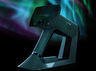
Optomed has spent the past two decades developing fundus camera technology that simplifies eye screenings for diabetics. The company is on the fifth generation of its camera system, known as Optomed Aurora.
The Aurora provides doctors with significant advantages over the direct ophthalmoscope and gives a clearer view than ever of the eye’s fundus. The camera features:
- 50-degree field of view, much wider than the ophthalmoscope
- Non-mydriatic operation, works without pupil dilation
- Autofocus and auto-exposure
- High contrast
The camera system is easily integrated into hospital IT systems. Images are captured in patient health records and are shareable over hospital systems for specialist review.
In addition, the Aurora can be integrated with AI software. This gives doctors access to various algorithms developed to help them assess signs of potential eye disease.
See the Future with Optomed
The Optomed Aurora is an ideal technological solution to provide capabilities for conducting quick, accurate retinal scans more widely. It is lightweight and easy to use, so doctors can take it from exam room to exam room and perform retinal scans at remote clinics and during house calls.
The Optomed Aurora is poised to play a role in protecting the sight of millions and changing the way that we detect eye disease.
Here at Optomed, our mission is to help save the vision of millions of people. By integrating our software and artificial intelligence solutions with our camera, we enable eye screening for everyone, wherever they are. To see how we can equip you to save the sight of more patients.
Share this insight