Technology is advancing quickly in the world of eye care. As devastating eye diseases continue to surge in the United States, the power of the newest technology can aid eye care professionals in the early detection of vision-threatening eye diseases.
According to the Center for Disease Control (CDC), diabetes in the United States now affects about 1 in every 10 people and more than 1 and 3 people have prediabetes. Many of these millions of people aren’t even aware their vision could be at risk for diabetic retinopathy.
While diabetes affects any age, another retinal disease is severely impacting our elderly population. Macular degeneration is a growing concern as the leading cause of blindness for seniors. Finally, the “silent sight seeker” shows that at least half of people with glaucoma aren’t even aware of this blinding eye disease.
These are only 3 of the main retinal diseases stealing the sight of the nation. They all have one important factor in common: they damage the retina. Viewing the retina allows eye care professionals to properly detect and manage their patient’s eye health.
In the United States, a 2017 study showed around 93 million people were considered at high risk for vision loss due to diabetes, eye problems, and being 65 years or older. Yet 40% did not see an eye doctor or get an eye exam. People need more comprehensive eye care screenings and eye care professionals need technology they can depend on.
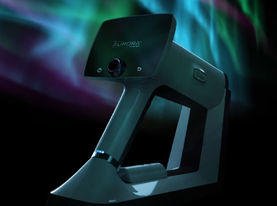
The Optomed Polaris provides eye care professionals with a fully automatic, non-mydriatic fundus camera with a small footprint to work in all types of clinics. This stationary device requires minimal training and provides sharp quality 45° images of the retina to improve the lives of patients and help eye care providers in the early detection of various retinal diseases.
The Importance of Using Fundus Images for Early Detection in Ocular Diseases
Studies continue to prove that early detection is key to preserving vision, yet many diabetics don’t get early screening. Guidelines for newly diagnosed diabetic patients recommend an eye exam within 6 months.
Studies show that, of patients with excellent access to health care, only about 40-50% make an appointment. Those with limited health care access drop to 20%. At best, only 50% of new diabetics get the eye screening they need to catch early signs of disease.
Early diagnosis and treatment of diabetic retinopathy decrease the risk of severe vision loss by 90%. By incorporating Optomed Polaris, more eye clinics can facilitate increased screenings and improved eye care for diabetic patients. If severe vision loss can decrease by 90% with early detection, more eye care professionals need to utilize available technological resources like fundus imaging.
The importance of effective detection tools applies to the other main eye diseases as well. With glaucoma as the leading cause of blindness among Hispanic and African Americans, early detection is critical. Medication easily treats both glaucoma and macular degeneration, so ophthalmologists and optometrists offer more people improved and autonomous lives with early detection. Using fundus imaging, eye care professionals can better view the optic nerve and macula, look for abnormalities, and diagnose these vision-threatening diseases before damage occurs.
Saving millions of patients’ vision is one huge benefit of the new Optomed Polaris camera. Additionally, fundus imaging offers a window into the overall health of the body. Detailed imaging of the retina gives eye care professionals more insight on conditions such as:
- Diabetes
- Hypertension
- Giant Cell Arteritis
- Lyme Disease
- Toxicities from medications
- And more…
Providing eye care professionals with medical devices that aid in early detection and appropriate treatment vitally supports their patients’ overall health and vision. Optomed Polaris helps eye care professionals better serve their patients.
Benefits and Features The Optomed Polaris Provides for Eye Care Professionals
Less Operating Time
Optomed Polaris aims to make the lives of eye care professionals easier. With automatic alignment and focus, staff or professionals won’t be spending unnecessary time adjusting the patient into the machine. The camera also has an adjustable chinrest that makes it easier for patients.
45° Views of The Retina
The 45° field of view is particularly helpful in diabetic retinopathy, as early diabetic changes can often be found in the periphery first. The camera provides 3D tracking and auto-capture to ensure quality images of the entire retina.
Close Monitoring
A key tool in fundus diagnostic imaging is follow-up pictures. This camera makes it easy to view prior images next to new ones with a 12MP sensory resolution and 10” touch screen for convenient navigation. This allows eye care professionals to make direct comparisons to prior images to assess changes in the retina.
Billable Images
Not only do patients benefit from early detection and treatment of various retinal diseases, but eye care professionals can bill for the images obtained. With more optometrists and ophthalmologists utilizing fundus imaging with Optomed Polaris, eye screenings will lead to greatly improved patient care.
Non-mydriatic
A fundus camera that doesn’t require dilation doesn’t interrupt clinic flow. This is a huge time saver since only some patients require dilation to view and assess retina health.
Small Footprint
Optometrists and Ophthalmologists won’t need an office build-out with the Optomed Polaris. The small footprint allows for use in every type of office setting. With the PC being integrated into the convenient touch screen of the device, all that’s needed is 11” by 19” of tabletop space.
Multiple Image Settings
Use the camera for both retinal and anterior segment images. Other key settings include red-free pictures to assess vessel detail, depth of pigmented lesions, and early detection of loss in the retinal nerve fiber layer.
Montage is another important tool to help eye professionals in the diagnostic and treatment of various retinal diseases. This is particularly helpful in diabetic retinopathy to assess the grading of dot/blot hemorrhages and vessel health.
Minimal Training
Luckily this device requires minimal training. The camera is easy to use for staff and patients with a proven high success rate in the first image captured. With 3-D tracking and the ability to auto-capture images in seconds, the camera helps save time for busy clinics and minimize patient interaction for this critical step in eye care.
AI Software and Integration
Optomed believes the future of retinal imaging will rely on AI technology. Optomed Polaris allows AI integration with any FDA-approved software.
AI integration allows for telemedicine, another growing component in the future of eye care. Optometrists are able to easily share images with ophthalmologists for timely referrals and treatment.
It’s time to put the Optomed Polaris in the hands of eye care professionals everywhere to increase retinal screenings and save vision.
Our mission is to help save the vision of millions of people. By integrating our software and artificial intelligence solutions with our camera, we enable eye screening for everyone, wherever they are.
