Artificial intelligence (AI) applied to medical imaging has shown great promise in recent years. In ophthalmology, there is particular interest in using AI algorithms to analyze fundus photographs to detect retinal diseases. These autonomous AI systems provide diagnostic recommendations directly, without physician interpretation. This allows the AI to perform specialized tasks previously only conducted by extensively trained eye care providers – who make up roughly 0.02% of the US population. Rigorously validated autonomous AI systems hold promise for improving access to care, efficiency, accuracy, and costs, while enabling physicians to focus their efforts on patients who need specialist management. However, autonomous AI systems assume full medical liability for their clinical decisions.
Early AI Results
Several studies have demonstrated high accuracy for AI systems compared to clinician grading for diseases like diabetic retinopathy (DR) and age-related macular degeneration. Diagnostic AI must have well-defined and disease-specific indications for use, which then need to be rigorously validated in preregistered studies for safety, efficacy, and equity, involving real-world workflow.
Over 90% sensitivity and specificity was observed for an AI model applied to a large fundus photograph dataset. AI screening for DR has even been approved by the FDA, with one product showing 87% sensitivity and 80% specificity in clinical validation studies.
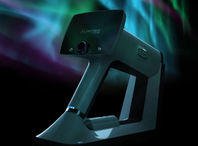
Several studies featuring the Optomed Aurora nonmydriatic handheld fundus camera have resulted in even more impressive numbers. Without dilating eyedrops and only a single macula-centered 50° photograph obtained with the autofocus and the auto exposure features the Aurora camera and AI were able to achieve 96.8% sensitivity and 96.8% specificity. There was also 100% agreement between the retina specialist using biomicroscopic examination and the blinded operator who classified the images obtained with the device. The Optomed Aurora currently has an FDA approved workflow using the AEYE Health algorithm with similar success rates. The Aurora AEYE has already been successfully implemented in DR screening programs.
Human Accuracy
Previous studies have shown that board-certified ophthalmologists that perform indirect ophthalmoscopy achieve between, to 91-specificity, and diagnosability for diabetic retinopathy. These are important safety measures as sensitivity and specificity are the measurements of false-negatives and false-positives within these patient trials, while diagnosability is important in what percent of patients get a valid result – either positive or negative. The ADA standards of care recommend retinal photography with remote reading or slit-lamp biomicroscopy by an eye care provider for retinal screening in diabetes. Neither the ADA nor the AMA specify required sensitivity/specificity metrics. Non-mydriatic photography paired with telehealth reading of photographs may demonstrate similar sensitivity and specificity (78-98%, 86-90%, respectively) to dilated fundus exam performed by an ophthalmologist (84-92%, 92-98%, respectively).
ICO/ADA guidelines state that adequate DR screening should include a visual acuity exam and retinal examination. Adequate visual acuity screening can be done with a refracted or presenting exam using an eye chart. Adequate retinal examination can be done via ophthalmoscopy, slit-lamp exam, or retinal photography (including 30° to wide field, mono or stereophotography, dilated or undilated). Retinal imaging and interpretation do not require a medical degree if proper training is completed.
Cost-Effectiveness of AI DR Screening
Beyond accuracy metrics, real-world implementation studies are examining the clinical workflow integration and cost-effectiveness of AI screening, especially DR. AI evaluation of fundus images in primary care offices increase adherence to follow-up eye care recommendations. Compared to in-office dilated eye exams, one study found a 23.3% cost reduction with its AI screening program. Meta-analyses of clinical trials with AI DR vs human graders showed that most AI screening programs are more economically efficient than traditional screening approaches for DR. While ongoing research is still needed, the evidence so far indicates AI-based DR screening can improve outcomes and reduce costs when incorporated thoughtfully into clinical practice.
The Big Picture
DR is a leading cause of vision loss worldwide. Early detection and treatment is critical, but implementing systematic DR screening is challenging due to resource constraints and complex coordination needed. Annual specialist exams are often unsustainable and insensitive compared to fundus photography. However, manual evaluation of all photos is time and resource intensive. Automated image analysis and AI algorithms can dramatically improve the efficiency and accessibility of teleophthalmology DR screening programs.
Handheld retinal cameras are a key technology for expanding DR screening, enabling image capture outside traditional tabletop settings. Recent studies have shown handheld cameras like the Optomed Aurora IQ can produce images of sufficient for accurate DR grading, even without pupillary dilation. Clinical and real-world studies have shown that Optomed Aurora IQ handheld fundus imaging is as effective as standard desktop fundus cameras in DR and diabetic maculopathy detection. The high sensitivity (91.8-96.9%) and specificity (94.8-100%) values for detecting the presence of any degree of DR, combined with the portability, and affordability, Optomed Aurora fundus imaging can improve population-level coverage.
However, challenges remain in applying AI successfully. Images must meet quality standards, training data should represent diverse populations, and clinicians require guidelines for evaluating model outputs. Well-designed prospective studies are needed to determine real-world clinical utility. But the existing evidence shows AI fundus analysis could expand access, support providers, and improve outcomes – something our stressed healthcare systems could benefit from.
Integrate the Aurora AEYE DR screening solution into your workflow today and join the cutting-edge.
Share this insight