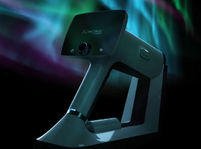
The FOTO-ED series of studies written by Bruce Beau et al. highlights the potential of incorporating nonmydriatic ocular fundus photography as part of routine emergency department (ED) care. They are seminal works and deserve their own in-depth discussion on the related studies, and what it means for clinic workflow and outcomes. The study’s objective was to examine the feasibility of non-mydriatic fundus photography as a promising clinical alternative to direct ophthalmoscopy in emergency departments.
The study’s importance lies in the fact that the ocular fundus is the only location in the human body where blood vessels can be directly observed, making it a valuable diagnostic tool for a variety of medical conditions. A fundus examination is crucial for identifying and treating many medical and neurological conditions. Unfortunately, direct ophthalmoscopy is often neglected and tricky to execute without using eye drops that dilate the pupils. To tackle this issue, the study examined the proposal that non-mydriatic ocular fundus photography (i.e., without dilating eye drops) could serve as a practical alternative to direct ophthalmoscopy. This is especially true in emergency departments, where doctors may have limited ophthalmoscopy training, a demanding schedule, and underappreciation of the value of eye exams, putting patients at risk for poor outcomes and doctors at risk for legal repercussions.
The FOTO-ED study comprises a two-phase sequential trial, intended to compare the routine use of direct ophthalmoscopy by emergency physicians (EPs) in phase I, with the routine use of nonmydriatic ocular fundus photography, interpreted by EPs, in phase II.
Phase I of the FOTO-ED study
The study ” Nonmydriatic Ocular Fundus Photography in the Emergency Department,” published in the New England Journal of Medicine, addresses the significance of nonmydriatic ocular fundus photography in the Emory University Emergency Department. 350 patients were enrolled in the trial who presented to the ED with a chief symptom would merit a fundus examination; headache, acute neurological deficit, acute vision loss, or diastolic blood pressure 120 mm Hg or higher. These patients were diagnosed and treated as standard of care by the ED physicians, while at the same time their fundus photos were reviewed by a neuro-ophthalmologist. The ED physicians were unaware of the photography results.
During the routine evaluation, ophthalmoscopy was only performed in 14% of the cases, with the ED physicians missing over 80% of previously unknown findings that were potentially relevant to acute patient management. Of the previously unknown relevant findings, 18% were identified by ophthalmologic consultation at the ED, and 82% were identified solely by fundus photography. Positive relevant findings included:
- optic nerve edema,
- intraocular hemorrhage
- hypertensive retinopathy
- arterial vascular occlusion
- optic nerve pallor
Most of these findings were only identified by the use of nonmydriatic fundus photography. Of these photographs, 97% were found to be of diagnostic value. And with an average time to photograph lasting 1.9 minutes, which is typically less than 0.5% of the patient’s total ED visit, brings potentially huge gains in efficiency for the organization. Remember, these photographs were taken by nonphysician staff.
Phase II of the FOTO-ED study
EPs in phase II were given immediate access to review the fundus photos in real time, without additional training. As in Phase I of the trial, nonmydriatic ocular fundus photography was interpreted by neuro-ophthalmologists and served as the reference standard. Compared with the first phase in which they only examined 14% of the patients by direct ophthalmoscopy, they examined 239 of 354 (68%) patients in phase II by using fundus photography, and reported that the images were helpful in 125 (35%) cases. The EPs were also better able to visualize pathology, and identified 16 of 35 relevant findings (46%). Emergency physicians used fundus photos much more frequently than they performed direct ophthalmoscopy, and their detection of relevant abnormal findings improved because of it. They also noted that it often improved care even when results were normal, being able to identify 96% of normal findings.
Case Study
Let’s consider a real case posed by the FOTO-ED study: A young woman complains of severe headaches and visits several physicians’ offices and emergency departments. Brain scans come back normal, and only a few doctors perform direct ophthalmoscopy, documenting that the optic disc was “difficult to visualize but likely normal.” Several weeks later, the patient goes to an ophthalmologist and is diagnosed with irreversible visual loss caused by chronic papilledema, leading to a medical malpractice lawsuit involving the earlier care providers.
Now, imagine a different scenario where the ED staff had taken photos of the patient’s retina and optic disc without the use of pharmacological dilation during the initial assessments. These images would have shown the papilledema clearly, leading to timely diagnosis and management, and avoiding the patient’s permanent vision loss.
Conclusions
The findings from these series of studies provide evidence that nonmydriatic ocular fundus photography is a feasible alternative that can be performed quickly and accurately in the ED setting. As we know, direct ophthalmoscopy is challenging, even in the most skilled hands. Even ophthalmologists miss about half of the relevant fundus findings with direct ophthalmoscopy compared with the indirect technique.
By providing a rapid and non-invasive means of evaluating ocular pathology, this technique has the potential to improve patient outcomes and expedite the diagnosis and treatment of ophthalmologic emergencies in the ED. We think it’s fair to posit that most EPs and ophthalmologists would welcome an alternate means of fundus evaluation in patients presenting with acute complaints, including headache and visual changes. The ED plays a critical role in the diagnosis and management of ophthalmologic emergencies, and fundus photography can provide valuable information for the evaluation of ocular pathology.
