Ophthalmologic exams in the outpatient and emergency room setting have enormous diagnostic value when performed consistently and correctly. In this article, we will briefly review:
- What happened to the fundoscopic exam?
- The latest published research in emergency and outpatient settings.
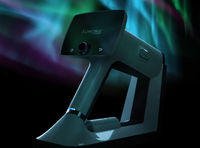
- Why the handheld Optomed Aurora IQ retinal camera will become the standard of care.
- A differential diagnosis list for papilledema (bookmark this for later).
The Antiquated Fundoscopic Exam
While the presence of papilledema can communicate a breadth of information regarding a patient’s status, rarely do providers properly identify it. Unfortunately, fundoscopy exams often fall to the wayside due to:
- Difficulty performing direct ophthalmoscope and limited field of view seen
- Patients inability to tolerate bright lights or dilation.
- Inability to document the findings in the EMR and share findings.
- Lack of ophthalmology services in rural settings.
In recent years, retinal photography has become increasingly available, with numerous studies suggesting that digital retinal photography will eventually replace traditional ophthalmoscope exams. The Optomed Aurora IQ can take high resolution images quickly, efficiently, and without pupillary dilation. Studies report that retinal photography has high sensitivity, specificity, and inter/intra-examination agreement when compared to in-person ophthalmologist examination.
Many new medical school and non-ophthalmology residency graduates perceive traditional fundoscopy exams to be burdensome and less useful than other methods. There is very little ophthalmology integrated into medical school curricula, with reports showing that ophthalmoscopy is performed infrequently and often inaccurately by medical students and residents.
The TOTeMS study (Teaching Ophthalmoscopy to Medical Students) aimed to compare the accuracy and preferences of medical students learning to examine the ocular fundus. Of 119 students, 92 (77%) preferred fundus photography over ophthalmoscopy, with that majority reporting more accurate assessments and less rates of frustration. Seventy percent indicated moving forward in their practice, they would prefer to use fundus photographs than traditional methods.
In a follow-up study performed one year later, these numbers remained consistent, with 76% still stating they prefer to use photographs. A cross-sectional, randomized, cross-over study of 146 second year medical students showed a statistically significant improvement (P < 0.001) in perceived usefulness and ease of use when using a camera over direct fundoscopy.
These results confidently predict that retinal cameras like the Optomed Aurora IQ will be the top preference for the next generation of physicians.
A recent article published in Neurology Clinical Practice called ocular fundoscopy a dying art. The authors encouraged preceptors on Neurology rotations to show medical students how to use retinal cameras to improve student confidence in interpretation of ocular fundus findings and improve awareness of the importance of performing ophthalmoscope exams.
Hidden Gems in the Emergency Department
According to phase I and phase II of the FOTO-ED study (fundus photography vs ophthalmoscopy trial outcomes in the emergency department), of the 350 patients evaluated in the emergency department with headache, focal neurological deficits, visual changes, or elevated diastolic blood pressure, 44 had relevant ocular fundus abnormalities (retinal hemorrhages, optic disc edema, hypertensive retinopathy, optic disc pallor, papilledema, etc). Alarmingly, many of those abnormal fundus exams had unremarkable physical exams and normal neuroimaging studies.
In the first phase of the study, 13% of patients had ocular fundus findings relevant to their ED management found by camera photography reviewed by neuro-ophthalmologists. Emergency Medicine physicians (EPs) missed every single notable ocular finding, who examined only 14% of enrolled patients by direct ophthalmoscopy.
This clearly demonstrated the critical role of ocular fundoscopy in patients in the ED with headache.
The FOTO-ED study also evaluated the time, ease, and quality of the photographs taken. Per neuro-ophthalmology, 87% of patients had a high-quality photograph, and mean ratings for ease, comfort, and speed by nurse practitioners and patients were ≥ 8.7 on a 10-point scale (10 best). Median photography session lasted 1.9 minutes, typically accounting for less than 0.5% of the patient’s total ED visit.
This study also demonstrated that non-mydriatic fundus photography is superior at detecting clinically important pathology when compared to direct ophthalmoscopy by EPs. Fundoscopy was well-received by patients and staff, and only required a trivial amount of patient’s time spent in ED.
In Phase II of FOTO-ED, EPs used non-mydriatic fundus photographs five times more frequently than they performed direct ophthalmoscopy, and their detection of relevant abnormalities improved substantially. Ocular fundus photography often assisted ED care even when normal, indicating that the ability to verify the absence of certain findings on ocular fundus examination was as important, if not more important, to ED clinical care situations than identifying abnormalities.
A pilot study published in the journal Acta Neurologica Scandinavica compared the results of a portable fundus camera to an ophthalmoscope. After enrolling 60 patients that presented to the ED with either a headache, cerebrovascular disorder, or in a state of delirium, fundus photography succeeded in 56 patients (93%) at detecting critical ophthalmic findings.
Optomed Aurora IQ Retinal Camera as the Standard of Care
Quickly identifying papilledema can lead to life and vision saving interventions. Even mild abnormalities are reported to communicate a wealth of knowledge to Neurologists; patients with TIA and stroke are more likely to have signs of microvascular retinal disease than the general population.
One study performed in Australian emergency departments found the prevalence of urgent fundus pathology to be as high as 16% when providers used non-mydriatic fundus photography (NMFP). The camera can easily be used on bed-bound patients.
A subsequent study showed that the use of NMFP influenced changes in management in 52 (39%) of 133 enrolled patients. Of these, 65% were escalations of management due to acute fundus pathology, while 35% were de-escalating changes following normal fundus findings. The diagnostic accuracy for acute fundus pathology improved from 0% to 29% in sensitivity and from 59% to 84% in specificity using DO and NMFP respectively.
Telemedicine and Remote Screening
The use of retinal cameras also shows great promise in telemedicine and remote screening. A retrospective study published in Telemed J E Health included 103 consecutive eyes with optic disc edema. The photographs were taken by members of an eye care clinic who had no prior ophthalmic photography training. When neuro-ophthalmologists later reviewed the images, they determined that the sensitivity and specificity for detection of optic disc edema were 71.8-92.2% and 81.6-95.2%. This satisfied the suggested requirements for new technologies that facilitate remote consultation (>80%) and is in line with reliability indices found in other telescreening studies.
A handheld, non-mydriatic camera is particularly well suited to settings where portability must be maximized and pupil dilation occurs rarely or cannot occur. The camera used in the aforementioned study can take high-quality photographs to detect optic disc edema with >80% sensitivity in patients whose pupils are not pharmacologically dilated. Training requirements are minimal, and a nonphysician can take photographs.
Papilledema Differential Diagnosis
Early detection and treatment of papilledema can notably reduce rates of blindness, but less than half of patients in the United States receive annual eye exams. Utilizing retinal cameras like the Optomed Aurora IQ in the primary care setting can help to alleviate this gap in care.
When performed correctly, identifying papilledema can help providers determine whether or not brain imaging is truly necessary, and thereby expedite diagnosing the following conditions:
Intracranial mass lesions (tumors, hematoma).
- Any form of cerebral edema, such as:
- Traumatic brain injury (TBI).
- Cerebral infarct.
- Acute hypoxic-ischemic encephalopathy.
- Increased CSF production (choroid plexus papilloma).
- Decreased CSF absorption, commonly seen after bacterial meningitis.
- Obstructive hydrocephalus, especially in pediatric cases.
- Venous sinus thrombosis (excellent physical exam maneuver to perform when debating whether or not to order that brain MRI on a patient in the ED).
- Acute complication of Sarcoidosis.
- Anterior Ischemic Optic Neuropathy (AION).
- Central Retinal Vein Occlusion (CRVO).
- Uveitis (autoimmune or infectious).
- Pseudopapilledema and optic nerve head drusen.
- Many other rare syndromes, diseases, and inflammatory disorders:
- Vogt-Koyanagi-Harada syndrome.
- AV fistula.
- Intraspinal lesion.
- Chronic respiratory illness.
- Toxic/metabolic conditions.
Routine fundoscopic exams have been recognized as a proven practice for identifying prognostic markers in numerous diseases:
- Diabetes (Proliferative diabetic retinopathy is one of the leading preventable causes of blindness).
- Hypertension
- Retinopathy of prematurity
- Glaucoma
Contact Optomed to Help
Working together with primary care, neuro-ophthalmologists, and emergency room personnel, Optomed aims to provide technology for the improved diagnosis and treatment of eye diseases, such as diabetic retinopathy and other conditions, with the goal of minimizing blindness.
Want to learn more about how we’re helping neurologists, neuro-ophthalmologists, and emergency staff?
Share this insight