Central retinal artery occlusion (CRAO) is an emergency situation. Diagnosis and treatment must be prompt to save a patient’s vision. The critical window of time for diagnosis and treatment is between four and six hours. Approximately 75% of those with CRAO will lose some sight in that eye. Risk factors of CRAO are the same as in strokes, so it’s important for the patient to monitor cardiovascular risk factors. When a patient comes to the emergency room (ER) and has symptoms that indicate CRAO, prompt diagnosis is important. Fundoscopy is the best way to diagnose the condition. However, most emergency rooms do not have access to fundus photography. Ophthalmoscopes are more common but can be difficult to use, and ER staff may miss subtle signs of retinal or optic disc changes. Ideally, an ophthalmologist would be on call to interpret any eye exams, but this is not feasible, especially in smaller hospitals. Tabletop fundus cameras are expensive and fixed to one place, making them out of reach of most hospitals. This is a problem because fundus photography is one of the quickest diagnosis techniques and can help patients in that crucial treatment window.
One of the best ways of using fundoscopy to detect a CRAO is with a mobile fundus camera. Its mobility means it can easily be brought to the ER and used whenever a CRAO is suspected or someone with its symptoms (sudden vision loss in one eye, blind spots in the visual field, and/or blurred vision) is brought in for treatment. If the condition is diagnosed sooner, there is a higher chance of saving the patient’s sight.
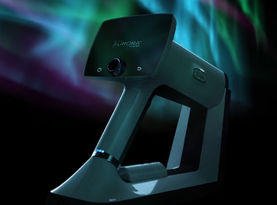
Nurse practitioners can also be trained in the technique, leaving more providers for other urgent care needs. The Aurora camera is one of the best ways of doing so, with an increased contrast-enhancing filter, a 50-degree field of view, and both autofocus and auto-exposure.
If the images provided by the mobile fundus camera are not enough to let the practitioners know of CRAO, there is also software that can be integrated to send those images to an ophthalmologist for further evaluation. For small hospitals that cannot afford to have a specialist on staff at all times, documenting with fundus photos gives them the security of knowing that they can always get a second opinion. For clinics that have them, artificial intelligence programs can also be integrated with the camera. The Aurora IQ can be easily integrated into any system without difficulty and can work with a variety of different software programs.
Like any other medical provider, ER staff wants to provide the best care to patients as quickly as possible. But if the department does not have the technology or medical personnel to render care for an acute condition, everyone suffers. Fundoscopy with mobile fundus cameras that can detect CRAO and are backed by telemedicine software that can send the pictures to ophthalmologists if needed can help the ER staff meet that need.
Patients experiencing CRAO need IV tPA within 4.5 hours for the best outcomes. That means you need to act FAST. Our mobile fundus cameras are easy to use for staff, enabling teams to easily confirm or refute CRAO in the ED. Digital image sharing helps emergency teams save critical time by streamlining communication.
Want to learn more about how we’re enabling neurologists, neuro-ophthalmologists, and emergency staff to confirm or refute CRAO faster and easier than ever?
Share this insight