Central retinal artery occlusion (CRAO) is a medical emergency, analogous to cerebral ischemic attack, and is classified as a stroke event. Recognizing the urgent nature of CRAO, the American Heart Association suggested in a June 2021 statement that the sudden, painless one-sided vision loss associated with CRAO should be communicated to the public as a potential sign of stroke, along with other widely recognized indicators, such as sudden, one-sided weakness, facial droop, and slurred speech. However, acute, monocular vision loss can be associated with multiple pathologies, so it’s vital to be able to quickly confirm or rule out a diagnosis of CRAO.
Prompt identification and treatment of CRAO can impact patients’ lives profoundly—in terms of vision, as well as stroke prevention—but it has been a challenge on numerous fronts, including a lack of specialized equipment and available ophthalmologic personnel in the emergency department. However, ED staff can now have access to a powerful tool in the movement to identify CRAO early and improve outcomes: the handheld mobile fundus camera.
Timely CRAO Treatment Translates to Better Outcomes
Several barriers to timely CRAO treatment have been identified in studies on the subject. In the emergency department in particular, a lack of specialized equipment and ophthalmologic personnel presents challenges. Overcoming these barriers to timely CRAO treatment is of paramount importance for several reasons.
First, prompt diagnosis can preserve the vision of patients with CRAO. If retinal blood flow is restored within ninety minutes, primate studies suggest that the chances are good that there will be no lasting damage. However, occlusions that aren’t resolved within four hours are shown to leave irreversible damage, which can greatly impact patients’ lives. For example, an increased risk of falling and functional dependence is associated with unilateral vision loss.
In addition to the high risk of irreversible vision loss, timely CRAO diagnosis can enable the identification of vascular risk factors that could lead to cerebral stroke or myocardial infarction. Patients are at the highest risk of stroke within the first seven days following CRAO, with up to 89% of these strokes being silent.
Lastly, timely and accurate identification and CRAO treatment better positions emergency providers in terms of risk management and medico-legal protection related to missed diagnoses and resultant poor outcomes.
Fundus Photography Can Aid in CRAO Diagnosis
Widespread agreement exists among neuro-ophthalmologic authorities that patients with acute monocular, painless vision loss should be examined immediately for signs of CRAO, including:
- Retinal pallor due to edema
- Cherry-red spot
- Vascular attenuation
- Boxcarring of the retinal blood vessels
Fundus photography imaging can easily detect these signs of CRAO, assisting in its diagnosis and distinguishing it from other disorders that can likewise cause sudden, painless monocular vision loss, such as vitreous and chorioretinal hemorrhage, retinal detachment, and acute optic neuropathy. This represents a real change in the ED, where those who lack on-site ophthalmology have been limited in diagnostic options in ocular emergencies.
In its June 2021 scientific statement on the management of CRAO, the American Heart Association noted that it is necessary to confirm the diagnosis of CRAO and rule out other disorders by using either a dilated funduscopic examination or a non-mydriatic color fundus photograph. According to this statement, if an ophthalmologist is not on site to confirm a diagnosis of CRAO, ocular fundus digital photography can be relayed to a specialist remotely.
Mobile Fundus Cameras Optimize Detection of CRAO in the ED
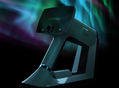
A leading manufacturer of handheld fundus cameras and technology worldwide, Optomed, has pioneered hardware and software solutions for various eye-screening needs. Its newest mobile fundus camera, the Optomed Aurora, is part of a revolution in CRAO detection in the emergency department.
The Aurora’s affordable price point removes cost as a barrier for most EDs to keep one on site. In addition, this camera is easily usable by ED staff, produces excellent image quality, and connects effortlessly with existing EHR software for remote specialist consultation.
Easily Usable by ED Staff
Emergency room physicians, nurses, and technicians can reliably obtain fundus photographs with minimal training. Research has demonstrated that non-mydriatic fundus photography can be reliably performed by non-physician emergency department staff in a short amount of time and that the practice is well-received by staff and patients in the ED.
The Optomed Aurora’s auto-focus and auto-exposure features enhance ease of use with minimal training. Its non-mydriatic operation translates to quick and easy examination, with no waiting on pupillary dilation. Its light weight and portability mean that it can easily be brought into patient rooms for examination.
Excellent Image Quality
The Aurora’s high-contrast, high-resolution imaging can detect even early-phase retinal changes, which can be instrumental in identifying anomalies, impairments, and retinal risk issues. Its 50-degree field of view provides an extensive look at the optical fundus. Images are more than adequate for detecting relevant findings for CRAO and other neuro-optical emergencies and disorders.
HIPAA-Compliant Connectivity
The Optomed Aurora is optimized to facilitate the quick diagnosis of CRAO, even in an ED setting with no on-site ophthalmology specialist. Digital images and videos can be documented and shared securely for remote consultation and easily integrated into existing hospital EHR systems.
A Handheld Fundus Camera Is a Powerful Tool for ED Staff
The immediate identification and treatment of CRAO have been recognized as essential in the preservation of vision and the prevention of further ischemic events that can drastically alter a patient’s life. ED staff members have had limited options in the past, but now they have access to technology that can powerfully impact outcomes in patients presenting with sudden, monocular vision loss. Handheld fundus cameras’ non-mydriatic operation, wide field of view, user-friendly design, high-quality digital imaging, and secure connectivity translate to timelier identification of CRAO, which in turn, can translate to better outcomes.
Patients experiencing CRAO need IV tPA within 4.5 hours for the best outcomes. That means you need to act FAST. Our mobile fundus cameras are easy to use for staff, enabling teams to easily confirm or refute CRAO in the ED. Digital image sharing helps emergency teams save critical time by streamlining communication.
Want to learn more about how we’re helping neurologists, neuro-ophthalmologists, and emergency staff?
Share this insight