Transforming the Diagnostic Process of Detecting and Treating Blinding Eye Disorders
Prevalence of Retinal Disease
The eye is a fascinating structure. Most of the time, it does an incredible job of delivering clear vision. However, that is not always the case. A wide variety of eye disorders can impact vision, from cataracts and glaucoma to retinal disorders. The most common retinal diseases are macular degeneration, diabetic retinopathy, and retinal vein occlusions. Other conditions that can impact the retina include retinal detachment and macular hole, among others.
Blindness in Retinal Disease
Diseases of the retina are essential to identify. They can have devastating consequences on vision. Many retinal conditions can lead to blindness if not caught early. The good news is that most of these eye disorders are treatable.
Approximately 11 million people in the United States suffer from macular degeneration. The prevalence worldwide is around 170 million. There are two types of macular degeneration: dry and wet. The dry form makes up approximately 90% of cases, while the wet form makes up the remaining 10%. It is vital to identify the dry form early, as the prescription of antioxidant vitamins can help slow its progression. Patients can also be counseled to look out for advancement to the wet form. They can be given specific tools to help with this. The wet form requires prompt diagnosis in order to coordinate treatment by a retina specialist.
In 2018, 34.2 million Americans, or 10.5% of the US population, had diabetes. The prevalence of diabetic retinopathy among these patients was 14.7%. In the early stages of diabetic retinopathy, patients need to be counseled on the importance of blood sugar control and the potential for vision loss. This is a crucial time for conversation, when it has the most potential for successful intervention. Without early treatment, more advanced forms of the disease, such as proliferative retinopathy or cystoid macular edema, can develop. These require treatment by a retina specialist.
Retinal vein occlusions are the second most common cause of vision loss due to retinal vascular disease. Although relatively rare, up to 0.2% for central vein occlusions and up to 2% for branched retinal vein occlusions, these conditions need to be identified and treated in order to prevent vision loss. Patients also need counseling on the systemic conditions that can contribute to retinal vein occlusions, such as hypertension, diabetes, and hypercholesterolemia. Again, time is of the essence when it comes to the diagnosis and treatment of these conditions.
The “Fundus Photography versus Ophthalmoscopy Outcomes in the Emergency Department” study found that 13% of patients who went to the emergency room with a headache, acute focal neurological deficit, acute visual changes, or diastolic blood pressure ≥120 mmHg have ocular fundus abnormalities. It is crucial that these be identified in a timely manner.
Retinal Examination
For the best treatment outcome, retinal diseases need to be diagnosed early. Although the American Optometric Association recommends annual eye examinations for those sixty-five and older, this does not always happen. Barriers such as time pressure, lack of access, insufficient education, fear, transportation problems, and insurance coverage issues prevent patients from getting eye exams.
The Primary Care Physicians’ Role in Detecting Eye Disorders
Although patients don’t always visit their eye doctor, they often do see their primary care physician. If a patient presents with a visual complaint, primary care physicians may want to use a direct ophthalmoscope to view the retina. However, this tool can be cumbersome and difficult to operate. The field of view is small, making it difficult to get perspective. The light needs to be sufficiently bright to properly view the retina, yet not so bright that patient cooperation plummets. The difficulties of using an ophthalmoscope can be discouraging, leading to avoidance of retinal exams and patient outcomes suffer as a result.
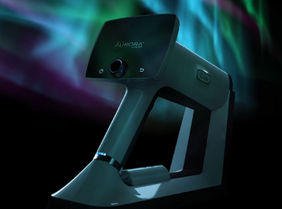
Optomed Aurora
A good quality non-mydriatic retinal camera can serve as an excellent substitute for direct ophthalmoscopy. It can provide more information on retinal health than was ever thought possible. Optomed Aurora is a portable handheld retinal camera that can be used in a primary care doctor’s office to screen for fundus abnormalities. It’s quick and easy, so it can be delegated to staff. It doesn’t require pupil dilation, which means patients can drive themselves home. The Optomed Aurora has a 50-degree field of view, compared to the 5-degree field of the direct ophthalmoscope. This enables fundus details to be sufficiently captured in a high-quality four-inch photograph. Images can then be read via telemedicine. Due to the camera’s clarity of imaging and consequent ease of interpretation, patients who need referrals can easily be identified and categorized by level of urgency. This technology can reach patients in rural and underserved areas, who may struggle with barriers such as transportation problems and lack of access to a specialist. Finally, there’s a tool that can identify retinal disease easily, allowing for prompt intervention and ultimately, preventing blindness.
Here at Optomed, our mission is to help save the vision of millions of people. By integrating our software and artificial intelligence solutions with our camera, we enable eye screening for everyone, wherever they are. To see how we can equip you to save the sight of more patients, schedule a free consultation today!
Share this insight