In 1851, when Hermann von Helmholtz introduced the ocular fundus exam using a direct ophthalmoscope, the practice of ocular medicine entered a golden age. Over the following decade, physicians were, for the first time, able to observe and describe in-vivo conditions, including central retinal artery occlusion, papilledema, and optic disc atrophy. Over the next century, ophthalmoscopy was incorporated into medical education and became a cornerstone of the general physical examination.
Direct Ophthalmoscopy Is Becoming Obsolete
In the last several decades, the limitations of direct ophthalmoscopy have been increasingly scrutinized. Mounting evidence reveals that direct ophthalmoscopy may be inadequately detecting conditions including diabetic retinopathy, glaucoma, and hypertensive retinopathy. This is true even when performed by experienced physicians, including ophthalmologists.
Attending Ophthalmologic Exams Can Be Challenging for Patients
There is concern regarding a noted lack of patient follow-through with referrals to ophthalmologic examination. Research has focused on exposing the reasoning behind this trend. Some patients possess limited awareness or misbeliefs surrounding the importance of screening. Costs associated with additional appointments are high financially and in terms of time and energy. Difficulty accessing screening centers can also contribute to the overall problem. Lastly, an array of emotional barriers exist for patients, including anxiety regarding complications of diabetes and the burden of disease management.
Moving Diabetic Retinopathy Screening into PCP Annual Wellness Checks May Be the Answer
More physicians are recognizing that accomplishing routine screening in the PCP office, perhaps with other routine wellness exams, can make a significant difference in meeting screening goals. It removes many significant barriers to adherence, including relying on patients to make arrangements to attend additional appointments, which may be cumbersome or challenging logistically, financially, or emotionally. With advances in technology over the past decade, high-quality digital screening of the fundus in a PCP’s office is both practical and effective.
As evidence of its efficacy has been mounting, leading scientists around the globe have been developing guidelines for successful and consistent practice of retina tele-screening for diabetic retinopathy. In February 2020, the Canadian Retina Research Network published their recommendations for Canadian national tele-screening standards, which are informed by similar programs already developed in England and Scotland.
Mobile Digital Screening Technology Is Reliable, Easy, and Cost-Effective
With modern handheld mobile non-mydriatic fundus cameras, reliable imagery can be captured easily, even by non-eye-care specialists.
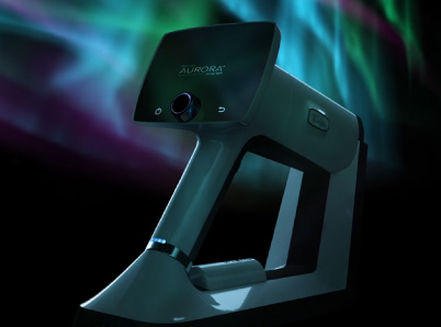
In 2013, the first handheld fundus camera to meet the ISO 10940 resolution requirements was developed by Finnish research and development company Optomed in pursuit of the goal to make vision-saving eye screening available to everyone. Since then, the technology has developed exponentially. Now in its fifth generation, Optomed’s Aurora offers a full 50-degree field of view, which is ideal for diabetic retinopathy screening. The Aurora is designed to consistently produce high-quality imagery with little investment of money, time, or training.
Reliable
Studies have shown that imagery captured using a mobile handheld fundus camera is adequate for confirmation or exclusion of various ocular and systemic conditions, including papilloedema, which can indicate intracranial conditions such as tumors, venous sinus thrombosis, CNS infection, hematoma, or intracranial hypertension.
A retrospective observational study, published in Ophthalmology Retina in April 2019, examined a teleretinal screening program in Texas in which 1,767 patients participated over a two-year period. The study concluded that teleretinal screening offered a high degree of accuracy in the detection of referable diabetic retinopathy. Another 2019 study of 978 Saudi patients with diabetes concluded that non-mydriatic fundus screening was practical and useful in the detection of diabetic retinopathy.
Easy to Use
Because it requires no eye dilation, this non-mydriatic camera is fast and easy to use in almost any setting. It has been designed for ease of use by non-eye-care professionals. Independent studies have demonstrated that even paraprofessionals can obtain reliable imagery with little training. The Optomed Aurora also connects seamlessly and securely to existing electronic medical record software.
Cost-Effective
The Optomed Aurora is available at an affordable price point, with minimal start-up costs and no installation or infrastructure costs. Little training is needed, so it can be put to use almost immediately.
Many PCP offices find that they can quickly recoup the initial investment costs by tapping into the diabetic retinopathy screening revenue stream.
Optomed Aurora Has Made Digital Screening and Fundus Eye Exams Easier for Primary Care Physicians
Optomed Aurora offers a practical, affordable, easy, and reliable method of optical digital screening right in the PCP’s office. This advanced technology enables PCPs to greatly enhance their patients’ outcomes for diabetic retinopathy and other ocular and neurological conditions.
Here at Optomed, our mission is to help save the vision of millions of people. By integrating our software and artificial intelligence solutions with our camera, we enable eye screening for everyone, wherever they are. To see how we can equip you to save the sight of more patients, schedule a free consultation today!
Share this insight